Micro-CT

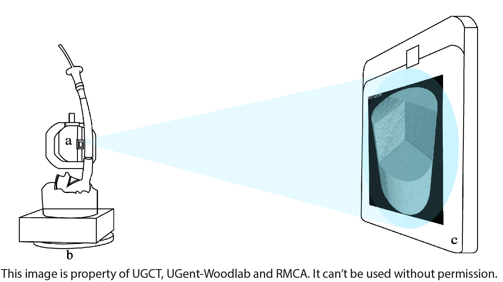
Bindi pipe (11,3 x 50,5 x 4 cm), scanned in the HECTOR scanner. The object is brought as close as possible to the X-ray source (a), using the rotation state (b), to capture a magnified, high resolution visualization of the internal wood structure on the detector (c). ®RMCA ®UGCT
X-ray Computed Tomography (CT), a non-invasive imaging technique, allows the internal structure of the wood inside objects to be visualized. A large selection of Congolese wooden objects are scanned at the UGent Centre for X-ray Tomography (UGCT — Ghent University (ugent.be)), using the High Energy CT scanner Optimized for Research (HECTOR). This micro-CT scanner was developed by the UGCT to be able to scan larger objects at high resolutions.
Below you can find two surface renderings of submicron X-ray CT scans that can be manipulated when pressing the play button.